Summary
The primary objective of this project is to achieve a profound understanding of the fundamental physical mechanisms that govern the behavior of damage in technological metal forming processes of steel. Capitalizing on this comprehensive knowledge of the underlying mechanisms, we will engage in collaborative endeavors with other projects focused on processes, characterization, and constitutive modeling. Our collective aim is to craft innovative design principles geared towards developing damage-tolerant forming processes and microstructures. During the first funding period, our emphasis was on developing an electron microscopy and A.I.-based methodology capable of characterizing the physical damage mechanisms in dual-phase steel DP800 at high resolution yet also large areas to bridge the scales from the nucleation of damage at the sub-micron scale to the variations of damage evolution in real microstructures and processes at the millimeter-scale. In the subsequent second funding period, we honed and tested the developed techniques, applying them to a different material, 16MnCrS5, and complex cases of damage characterization in three dimensions as well as under conditions of combined loading modes. In the final third funding period, our attention will be directed towards a detailed analysis of damage mechanisms and evolution in three dimensions and the intricate interplay between damage characteristics and microstructure. We will also delve into the role of big data and uncertainties in the methodologies used for damage characterization. Moreover, we aspire to advance current analysis techniques by integrating super-resolution scanning electron microscopy imaging to enable efficient quantification of damage over representative areas spanning over many square millimeters.
Our goals for the third funding period are:
- To establish a scale-bridging understanding of damage mechanisms in 2D and 3D.
- To highlight and explain differences in damage quantification in 2D and 3D
- To unravel the interaction between damage evolution and microstructure both in terms of local hardening by hard phases and local softening by recrystallization.
- To quantify and establish the role of uncertainties in characterization of damage and material properties.
- To develop methods for efficient imaging and analysis of damage in representative, large areas.
From the results and methods obtained within project B02 we expect to push the boundaries of damage characterization in steels, offering comprehensive insights into operating damage mechanisms across larger material areas and facilitating the integration of this knowledge into predictive models. This can guide material design and processing strategies to mitigate the formation of damage during forming processes.
Project progress to date
Initial objective and scientific state of the art
An in-depth understanding of damage processes, especially damage nucleation and growth, is indispensable to damage controlled forming processes. The separation of damage mechanisms and stress states during these processes has already proven fruitful in controlling the global damage during the first funding period. Because experiments focused on simple model processes at that time, such as tensile deformation and bending, the overarching objective of this project during the second funding period was to deepen the physical understanding of damage mechanisms and evolution. The foundations laid in the first funding period by the developed methods and results were expanded upon in a way that described the damage behavior of more complex samples with a wider variety, transitioning to circumstances that are closer to industrial processes. In particular, the developed method using scanning electron microscopy (SEM) panoramic imaging and machine learning-assisted automated analysis was applied to new materials and loading scenarios. This included a case-hardening steel 16MnCrS5 with different damage initiation and evolution behavior, as compared to the DP800 steel, which was analyzed in the first funding period. Other dual phase steels with different microstructures received from project B06 were also investigated. The efficient machine learning-based damage analysis was extended in terms of its application to include a 3D analysis of deformed microstructures, enabling tracking and classification of individual damage sites in all three spatial dimensions. Analysis of simple strain paths was also expanded upon in the second funding period by introducing more complex loading scenarios, comprising two-step strain path combinations of tension and bending. Additionally, the mechanical properties and active slip systems of non-metallic MnS inclusions and the surrounding ferrite matrix in 16MnCrS6 steel, responsible for the initiation of damage during forming were measured at ambient and elevated temperature. The most relevant results and the applied methods are described below in detail.
3D damage characterization in DP800
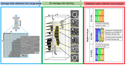
One achievement reached during the second funding period was the successful development and application of a machine learning assisted damage characterization method that allows the semi-automatic classification of the damage mechanism and damage site morphology, distribution and size in all three spatial dimensions. This methodology was developed by enhancing the automatic damage classification developed in the first funding period and employed panoramic scanning electron microscopy of the same sample surface at different depths achieved by metallographic serial sectioning followed by deep-learning assisted automated image analysis as shown in Fig. 1 [Med23a]. This method was applied to the DP800 dual-phase steel using ten slices of panoramic electron micrographs per sample separated by 0.25 - 0.5 µm intervals into the material depth. Through careful image registration and automated analysis of damage mechanisms using convolutional neural networks developed in the first funding period, the behavior and evolution of damage for thousands of sites was tracked in three dimensions. This method was used on tensile test samples with strains of 14%, 19% and 25%. The results of this work showed that, for the investigated microstructure, martensite cracking is the most dominant damage nucleation mechanism and the dominance of interface decohesion at higher strains is related to damage evolution and as such overestimated in 2D analyses [Med23a]. This highlights that classifying damage mechanisms solely from a 2D view lead to misleading results [Med23a]. However, it remains unclear how large the difference between 2D results and 3D results is and whether it may be eliminated. Furthermore, the 3D analysis revealed a strong dependence of damage density on metallographic preparation, exemplifying the need for standard and good practice metallographic procedures developed within TRR 188. The successful application of this method for 3D damage characterization allows a more in-depth look at the initiation of damage sites and can be used especially for further investigations of the effect of the local microstructure on damage initiation and to inform statistical damage models considering void morphology. The transfer between such big experimental data and computational modelling has further been supported by enabling phase segmentation across the large panoramic images, which is not possible by simple grayscale thresholding [Med24].
 |
|
Fig. 1: Workflow of 3D damage site analysis on SEM panoramic images. Damage sites are detected and tracked in 3D to extract statistical data about damage initiation mechanisms at different strains [Med23a]. |
Damage behavior of DP800 under complex strain paths
In the first funding period, damage analysis in the DP800 steel was mainly focused on simple model strain paths, used to successfully clarify the influence of different triaxialities on the damage behavior. To determine how damage occurs in samples that have been deformed in a way more similar to industrially relevant processes and to unravel the influence on strain path dependent properties, the damage behavior of two-step processes was analyzed in [Wol23b]. To this end, the already well-established processes of tensile and bending tests were compared to their possible combinations, leading to the following possible strain paths during forming: positive triaxiality (tensile area of simple bending before and after a tensile test), a combination of positive triaxiality after further positive triaxiality (bending after tensile test, tensile test after bending), positive triaxiality after negative triaxiality (tensile test after bending at compression side) and negative triaxiality after positive triaxiality (at compression side of bending after tensile test). The damage distribution in terms of the normalized number of voids is shown in Fig. 2 for all samples. The results reaffirmed that a high positive triaxiality leads to a growth of damage sites, as larger voids were observed for strain path combinations with higher triaxiality.
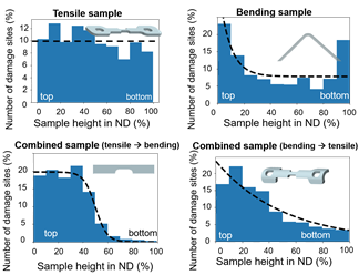 |
| Fig. 2: Comparison of the damage distribution with regard to the normalized number of voids along the sample thickness in normal direction (ND) for simple and combined strain paths. Schematic fits are included at the shown distributions to indicate the average damage behavior [Wol23b]. |
These experiments also highlighted the importance of the change of loading direction, strain hardening and damage prevalence from previous loading. The higher the number of damage sites from a previous deformation experiment, the higher the number of damage sites after a second deformation experiment, when deforming with a positive triaxiality. Changing the loading direction between two different deformation steps leads to less pronounced growth of voids as voids will nucleate in new positions.
Finally, deforming the material at negative triaxialities, resulting in a strain hardened microstructure and no damage formation, will decrease the damage formation in further deformation steps, due to the decreased mechanical contrast in the microstructure. This behavior can be observed in Fig. 3, highlighting the damage decrease in terms of void area fraction for the sample that was previously strain hardened by compressive stresses during bending.
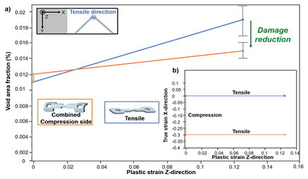 |
| Fig. 3: Change of void area fraction during tensile deformation in Z-direction for a simple tensile sample (blue) and the compression side of a sample that has been bent first in a). The strain path of both samples is given in b) [Wol23b]. |
Based on these observations, rules for two-step forming processes were derived: Firstly, forming processes should be structured in a way that they incorporate a change of loading direction after the material was formed with a positive triaxiality. Secondly, forming processes should start with forming steps that introduce no damage to the microstructure, but lead to strain hardening. Lastly, the number of damage sites from previous forming steps should be kept at a minimum.
Micromechanical characterization of different DP microstructures
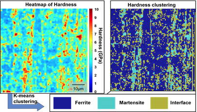
Quantifying the strain hardening behavior is essential, as it plays a critical role in damage formation. We could show-case this combining macroscopic tensile and microscopic compression testing [Tia24] and also for multi-step straining processes as described above, where a nanoindentation method was applied [Wol23b]. This method uses rapid indentation mappings to measure the strain hardening in different areas of the bending sample. This method separates the microstructural components martensite and ferrite based on their hardness and indentation modulus. A hardness map obtained from the center of a DP800 bending sample is shown in Fig. 4 next to the final clustered result, visualizing the mechanical contrast between martensite and ferrite.
 |
|
Fig. 4: Microstructural segmentation of DP800 microstructure into ferrite, martensite and a class of interfaces using hardness maps generated by rapid nanoindentation mapping. [Wol23b]. |
This method allows rapid discrimination of phases with high mechanical contrast even at small grain sizes and was also applied to different microstructures developed in project B06. Here, the mechanical properties of martensite and ferrite with different carbon contents and different morphologies were measured in bending samples. By comparing deformed and undeformed areas of the bending samples, the strain hardening behavior of the ferrite was also determined in these microstructures. By mapping areas in which damage initiation took place combined with imaging at different acceleration voltages, it was also shown that this method can detect sub-surface damage sites in the microstructure due the decrease of indentation modulus at places where subsurface damage sites are present. Additionally, this method provides a high data acquisition rate, as each indent takes about a second. The large data sets harbor the potential to be analyzed even more thoroughly to quantify the uncertainty of these tests, leading to better information about the mechanical properties of individual phases.
Method transfer: automated damage classification and quantification for 16MnCrS5 steel
To gather information about damage mechanisms and prevalence in a time efficient way that it is not limited to the DP800 steel, a computer vision machine learning workflow was devised and trained that classifies and measures damage in the 16MnCrS5 case-hardening steel. As damage initiation in this steel is related to the prevalent MnS inclusions found in the microstructure, the workflow shown in Fig. 5 [Wol23a] differs from that devised for the DP800 steel developed in the first funding period. Two machine learning networks are trained with two distinct purposes. The first network localizes and captures MnS inclusions inside of high-resolution panoramic SEM images. Once the second network is provided with these images, it localizes damage sites in the inclusions and if damage sites are found it classifies them according to the underlying mechanism as crack or delamination. After classification, the second network transfers images of the damage site to an algorithm that uses grayscale thresholding to separate pixels that belong to the damage site from pixels of the inclusion or the matrix, enabling the measurement of size and morphology for each damage site. Because of the two-step detection it is not only possible to assign damage parameters, such as void area fraction and number of voids, to the complete sample by summing up information about all damage sites, but damage parameters can be assigned to individual inclusions, which tend to fracture in multiple places. Applying this network to images taken during an in-situ tensile test made it clear that damage initiation does not happen simultaneously for all inclusions [Wol23a]. During these experiments it was shown that the local microstructure surrounding the inclusions, as well as the orientation of the inclusions in relation to the loading direction, has the potential to influence the damage prevalence and mechanism.
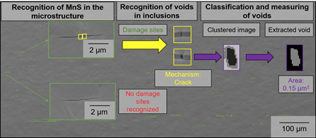 |
| Fig. 5: Workflow of machine learning assisted, automated damage recognition, classification and quantification in the 16MnCrS5 case-hardening steel [Wol23a]. |
In addition to the in-situ tensile test at ambient temperature, this method has also been applied to tensile test samples that have been deformed, but not to failure, at temperatures of 200°C, 400°C and 600°C. The characterization results showed that damage prevalence does not follow a linear trend with temperature but assumed the lowest damage prevalence at 400°C, before rising again at 600°C. Because the damage prevalence largely relies on the mechanical contrast of MnS inclusions and the surrounding matrix, additional micromechanical experiments at elevated temperatures were conducted on MnS.
Constitutive models for MnS inclusions
In order to enable modelling of damage formation during forming, it is essential to achieve a realistic crystal plasticity model for the MnS inclusions in the 16MnCrS5 steel. For room temperature, this was achieved in the first funding period [Kus20] and extended towards high temperatures in the second funding period. As the MnS inclusions are too small to be tested inside the 16MnCrS5 microstructure (cf. Fig. 5) and the indenter tips used for compression tend to chemically react with or are absorbed into iron, a MnS sample was prepared by heating MnS powder to melting temperature and letting it solidify. This provided grains large enough to fabricate several micropillars with identical orientations. In total, micropillar compression tests were conducted on 2 different orientations, each facilitating slip on one of the two common slip systems, and at 4 different temperatures (20°C – 600°C) spanning 40 successfully compressed pillars in total. From these experiments, the dependence of the yield strength and critical resolved shear stress of MnS on temperature was deduced, as calculated from compression curves such as shown in Fig. 6.
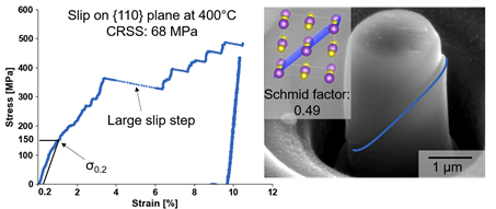 |
| Fig. 6: Compression engineering stress-strain curve of micropillar oriented for slip on {110} planes at 400°C and the corresponding micropillar after compression with highlighted slip steps next to a visualization of the crystal orientation of the micropillar, as well as the determined active slip plane. |
Project- and subject-related list of publications
| [Kus19a] | Kusche, C.F., Reclik, T., Freund, M., Al-Samman, T., Kerzel, U., Korte-Kerzel, S., 2019, Large-area, high-resolution characterisation and classification of damage mechanisms in dual-phase steel using deep learning. PloS one, 14(5), DOI: 10.1371/journal.pone.0216493. |
| [Kus19b] | Kusche, C.F., Dunlap, A., Pütz, F., Tian, C., Kirchlechner, C., Aretz, A., Schwedt, A., Münstermann, S., Al-Samman, T., Korte-Kerzel, S., 2019, Efficient characterization tools for deformation-induced damage at different scales. Production Engineering, 14, 95-104, DOI: 10.1007/s11740-019-00936-w |
| [Kus20] | Kusche, C.F., Gibson, J. S. K.-L., Wollenweber, M. A., Korte-Kerzel, S., 2020, On the mechanical properties and deformation mechanisms of manganese sulphide inclusions. Materials & Design, 193, 108801, DOI: 10.1016/j.matdes.2020.108801 |
| [Kus21] | Kusche, C. F., Pütz, F., Münstermann, S., Al-Samman, T., Korte-Kerzel, S. (2021), On the effect of strain and triaxiality on void evolution in a heterogeneous microstructure–A statistical and single void study of damage in DP800 steel, Materials Science and Engineering: A, 799, 140332, DOI: 10.1016/j.msea.2020.140332 |
| [Lin23] | Lin, B., Medghalchi, S., Korte-Kerzel, S., Xu, B., 2023, A Machine Learning Enabled Image-data-driven End-to-end Mechanical Field Predictor For Dual-Phase Steel. Proceedings in Applied Mathematics and Mechanics, 22(1), DOI: 10.1002/pamm.202200110 |
| [Med20] | Medghalchi, S., Kusche, C. F., Karimi, E., Kerzel, U., Korte-Kerzel, S., 2020, Damage analysis in dual-phase steel using deep learning: transfer from uniaxial to biaxial straining conditions by image data augmentation. JOM, 72, 4420-4430, DOI: 10.1007/s11837-020-04404-0 |
| [Med23a] | Medghalchi, S., Karimi, E., Lee, S., Berkels, B., Kerzel, U., Korte-Kerzel, S., 2023, Three-dimensional characterisation of deformation-induced damage in dual phase steel using deep learning, Materials & Design 232, 112108, DOI: 10.1016/j.matdes.2023.112108 |
| [Med23b] | Medghalchi, S., Zubair, M., Karimi, E., Sandlöbes-Haut, S., Kerzel, U., Korte-Kerzel, S., 2023, Determination of the Rate Dependence of Damage Formation in Metallic‐Intermetallic Mg–Al–Ca Composites at Elevated Temperature using Panoramic Image Analysis, Advanced Engineering Materials, 25(21), 2300956, DOI: 10.1002/adem.202300956 |
| [Med24] | Medghalchi, S., Kortmann, J., Lee, S.-H., Karimi, E., Kerzel, U., Korte-Kerzel, S., 2024, Automated Segmentation of Large Image Datasets using Artificial Intelligence for Microstructure Characterisation, Damage Analysis and High-Throughput Modelling Input, arXiv cond-mat, DOI: 10.48550/arXiv.2401.01147 |
| [See23] | Seehaus, M., Lee, S.H., Stollenwerk, T., Wheeler, J.M. and Korte-Kerzel, S., 2023. Estimation of directional single crystal elastic properties from nano-indentation by correlation with EBSD and first-principle calculations. Materials & Design, 234, p.112296. DOI: 10.1016/j.matdes.2023.112296 |
| [Tia24] | Tian, C., Kusche, C. F., Medina, A., Lee, S., Wollenweber, M. A., Pippan, R., Korte-Kerzel, S., Kirchlechner C., 2024, Understanding the damage initiation and growth mechanisms of two DP800 dual phase grades. Materials & Design, 112630, DOI: 10.1016/j.matdes.2024.112630 |
| [Wol23a] | Wollenweber, M.A., Kusche, C.F., Al-Samman, T., Korte-Kerzel, S., 2023, On the automated characterisation of inclusion-induced damage in 16MnCrS5 case-hardening steel, Advances in Industrial and Manufacturing Engineering, 100123, DOI: 10.1016/j.aime.2023.100123 |
| [Wol23b] | Wollenweber, M.A., Medghalchi, S., Guimarães, L.R., Lohrey, N., Kusche, C.F., Kerzel, U., Al-Samman, T., Korte-Kerzel, S., 2023, On the damage behaviour in dual-phase DP800 steel deformed in single and combined strain paths, Materials & Design 231, 112016, DOI: 10.1016/j.matdes.2023.112016 |